What Is Karyotyping, and Why Do Scientists Check a Stem Cell's Chromosomes Before It's Used in Therapy?
What this article covers
- What This Article Covers
- Before any stem cell line can be used in research or considered for a clinical trial, scientists have to answer a basic but critical question: has the cell changed in ways that make it unsafe or unreliable? One of the oldest and still most widely used tools for answering that question is karyotyping — a method for photographing and counting a cell's chromosomes to check that nothing important has gone missing, duplicated, or rearranged.
- What Is a Karyotype, Exactly?
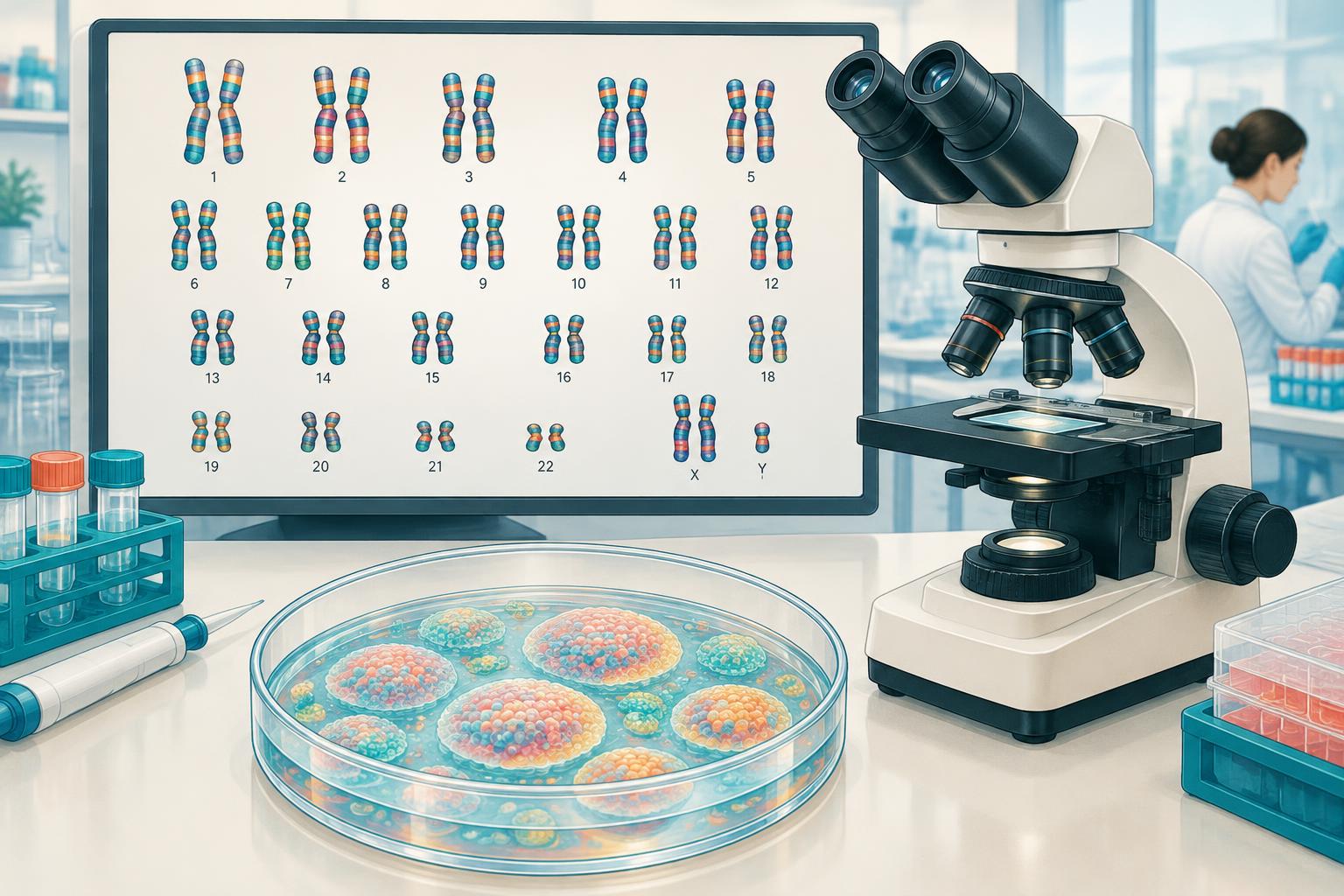
- Every normal human cell contains 46 chromosomes — 23 pairs, including one pair of sex chromosomes — bundled tightly inside the nucleus. A karyotype is essentially a sorted photograph of those chromosomes, arranged by size and pattern so a trained analyst (or software) can count them and compare their banding pattern to a known reference map.
- Why Stem Cells Are Prone to Chromosomal Change
- Unlike most cells in the body, stem cells intended for research or therapy are grown and expanded in the laboratory over many rounds of division — a process called passaging. Each time a cell divides, there is a small chance that errors creep into how DNA is copied or how chromosomes are separated.
- How Often, and When, Should Cells Be Checked?
- Because genetic drift is a moving target, responsible stem cell science treats karyotyping as a recurring checkpoint rather than a one-time stamp of approval.
- What Karyotyping Can — and Can't — Catch
- Karyotyping is powerful for what it does well: spotting large-scale problems, such as an entire extra or missing chromosome (aneuploidy) or a large chunk of one chromosome relocated to another (a translocation). But it has a real resolution limit.
What This Article Covers
Before any stem cell line can be used in research or considered for a clinical trial, scientists have to answer a basic but critical question: has the cell changed in ways that make it unsafe or unreliable? One of the oldest and still most widely used tools for answering that question is karyotyping — a method for photographing and counting a cell's chromosomes to check that nothing important has gone missing, duplicated, or rearranged. This article explains what karyotyping is, why stem cells are especially prone to developing chromosomal changes during laboratory growth, what a normal versus abnormal result looks like, and why this unglamorous quality-control step is one of the quiet foundations that responsible stem cell science depends on.
What Is a Karyotype, Exactly?
Every normal human cell contains 46 chromosomes — 23 pairs, including one pair of sex chromosomes — bundled tightly inside the nucleus. A karyotype is essentially a sorted photograph of those chromosomes, arranged by size and pattern so a trained analyst (or software) can count them and compare their banding pattern to a known reference map.
To create one, scientists stop dividing cells at a specific point in the cell cycle, stain the chromosomes with a dye that creates a distinctive light-and-dark banding pattern (a technique called G-banding), and then image and arrange them under a microscope. The result shows, at a glance, whether a cell has:
- The right number of chromosomes (46, not 45 or 47)
- Chromosomes of the expected size and shape
- No obvious large-scale rearrangements, such as a piece of one chromosome attached to another
Why Stem Cells Are Prone to Chromosomal Change
Unlike most cells in the body, stem cells intended for research or therapy are grown and expanded in the laboratory over many rounds of division — a process called passaging. Each time a cell divides, there is a small chance that errors creep into how DNA is copied or how chromosomes are separated. Over dozens of passages, these errors can accumulate.
Pluripotent stem cells (embryonic stem cells and induced pluripotent stem cells, or iPSCs) are particularly susceptible to this because of a phenomenon researchers call "culture adaptation." If a random genetic change happens to give a cell a growth advantage — for example, by nudging it closer to a cancer-like proliferation state — that cell and its descendants can quietly outcompete and overtake the rest of the dish, even though no one added anything to cause it. According to the International Society for Stem Cell Research (ISSCR), gains of chromosomes 1, 12, 17, 20, and X, along with mutations in the cancer-related gene TP53, are among the most commonly recurring culture-acquired changes seen across pluripotent stem cell lines worldwide. The ISSCR also notes that a cell population carrying such an advantage can come to dominate a culture within as few as five to ten passages, which is part of why ongoing monitoring — not a single one-time check — is the standard.
How Often, and When, Should Cells Be Checked?
Because genetic drift is a moving target, responsible stem cell science treats karyotyping as a recurring checkpoint rather than a one-time stamp of approval. ISSCR guidance recommends testing:
- Master and working cell banks, ideally right after a genetic bottleneck such as cloning a single cell or performing gene editing
- Roughly every 10 passages during ongoing culture, adjusted for how quickly a given cell line divides
- After major culture changes, such as switching media formulations, reprogramming methods, or any complex genetic manipulation
- At the end of an experiment or production run, to confirm the cells used were the same as the cells characterized
This matters just as much — arguably more — in manufacturing a cell therapy product intended for people, where a master cell bank and working cell bank are karyotyped and re-tested at multiple points to document genomic stability before any batch is released for use.
What Karyotyping Can — and Can't — Catch
Karyotyping is powerful for what it does well: spotting large-scale problems, such as an entire extra or missing chromosome (aneuploidy) or a large chunk of one chromosome relocated to another (a translocation). But it has a real resolution limit. Standard G-banded karyotyping can reliably detect changes only down to roughly 5 to 10 megabases of DNA — meaning a smaller duplication, deletion, or point mutation affecting even an important single gene can look completely normal on a karyotype.
This is why karyotyping is increasingly paired with higher-resolution techniques, including:
- Chromosomal microarray analysis, which can detect much smaller gains and losses of DNA
- Digital PCR or targeted sequencing for specific "hotspot" mutations, such as in TP53
- Whole-genome or whole-exome sequencing, used in research settings and increasingly in manufacturing, to catch changes invisible to a microscope
No single test catches everything, which is why the ISSCR and most regulatory frameworks encourage combining a broad structural check like karyotyping with a targeted molecular check for known problem genes.
Why This Matters for Safety, Not Just Research Accuracy
A chromosomal abnormality in a lab dish isn't automatically dangerous — some changes are neutral or specific to artificial culture conditions and don't survive in a living body. But some of the same changes recurrently seen in cultured pluripotent stem cells, like TP53 disruption, are also found in human cancers, which is precisely why regulators treat genomic stability testing as a non-negotiable safety checkpoint rather than a research nicety. For any cell therapy product, demonstrating that the manufactured cells haven't drifted into a genetically abnormal, potentially tumor-prone state is a basic requirement for moving toward human use — it's part of the broader manufacturing quality-control picture that also includes sterility testing, identity testing, and potency assays.
What This Means If You're Researching a Clinical Trial
If you're evaluating a legitimate stem cell clinical trial, genomic/chromosomal stability testing of the cell product is something you can reasonably expect to be part of the manufacturing and quality-control process described in trial documentation or publications — it's one small but meaningful signal, among many, that a program is being run with rigorous laboratory oversight rather than minimal characterization.
Bottom Line
Karyotyping is a decades-old, low-tech-sounding but genuinely essential tool for keeping stem cell science honest: it lets researchers verify that the cells they're studying — or manufacturing for a therapy — haven't quietly accumulated chromosomal changes during laboratory growth. Because pluripotent stem cells are particularly prone to this kind of "culture adaptation," leading scientific bodies recommend checking cell lines repeatedly throughout their life in the lab, not just once. Karyotyping has real limits — it can miss small but potentially important genetic changes — which is why it's typically used alongside more sensitive molecular tests. Understanding this quality-control layer helps put a stem cell product's manufacturing claims into proper context: genomic stability testing is a marker of rigor, not a guarantee, and it's one of many checkpoints a responsible program should be able to describe.
Key Questions Answered
- What does karyotyping check?
- It checks chromosome number, size, shape and large-scale rearrangements by imaging and sorting a cell's chromosomes.
- How often should stem cell chromosomes be checked?
- ISSCR guidance recommends recurring checks of cell banks, roughly every 10 passages, after major culture changes and at the end of an experiment or production run.
- Does a normal karyotype guarantee safety?
- No. Standard G-banding can miss changes smaller than roughly 5–10 megabases and important gene mutations. Genomic stability testing needs complementary molecular tests and is only one part of product quality control.
Sources
- International Society for Stem Cell Research (ISSCR), "Section 3: Genomic Characterization," ISSCR Standards for Human Stem Cell Use in Research, 2023, https://www.isscr.org/basic-research-standards/genomic-characterization
- Vales et al., "Gains of 20q11.21 in human pluripotent stem cells: Insights from cancer research," Stem Cell Reports / PMC, 2024, https://pmc.ncbi.nlm.nih.gov/articles/PMC10828824/
- "Longitudinal analysis of genetic and epigenetic changes in human pluripotent stem cells in the landscape of culture-induced abnormality," Frontiers in Cell and Developmental Biology / PMC, 2025, https://pmc.ncbi.nlm.nih.gov/articles/PMC11612254/
- AATBio, "What are the limitations of karyotyping?", https://www.aatbio.com/resources/faq-frequently-asked-questions/what-are-the-limitations-of-karyotyping
- U.S. Food and Drug Administration, "Chemistry, Manufacturing, and Control (CMC) Information for Human Gene Therapy Investigational New Drug Applications," https://www.fda.gov/media/73624/download
- Thermo Fisher Scientific, "Karyotyping Versus Chromosomal Microarrays: Detecting Chromosomal Abnormalities," https://www.thermofisher.cn/blog/behindthebench/karyotyping-versus-chromosomal-microarrays-detecting-chromosomal-abnormalities
Related Articles
- Fundamentals
What Is Angiogenesis, and Why Does It Matter for Stem Cell Therapy?
New blood vessels don't just show up — they're built, through a process called angiogenesis. Here's what it is, why the body depends on it for healing, and why so much stem cell research is focused on helping it along.
- Fundamentals
Myth: If a Treatment Is Listed on ClinicalTrials.gov, It Must Be Safe and Effective
Seeing a treatment listed on ClinicalTrials.gov can feel like a stamp of approval — but registering a trial costs nothing, requires no proof the treatment works, and isn't vetted by the government for safety or scientific quality.
- Fundamentals
What Are Organoids, and How Are They Grown From Stem Cells?
Organoids are tiny, self-organizing models grown from stem cells. They help study human biology and drug responses but are not complete transplant-ready organs.