What Are Stem Cell Surface Markers, and How Do Scientists Identify a Stem Cell?
What this article covers
- What a Surface Marker Actually Is
- Every cell is studded with proteins embedded in its outer membrane, many of which project outward where they can be detected from outside the cell. Which of these proteins a cell displays, and in what combination, depends heavily on what kind of cell it is and what state it's in.
- How Scientists Actually Detect Them: Flow Cytometry
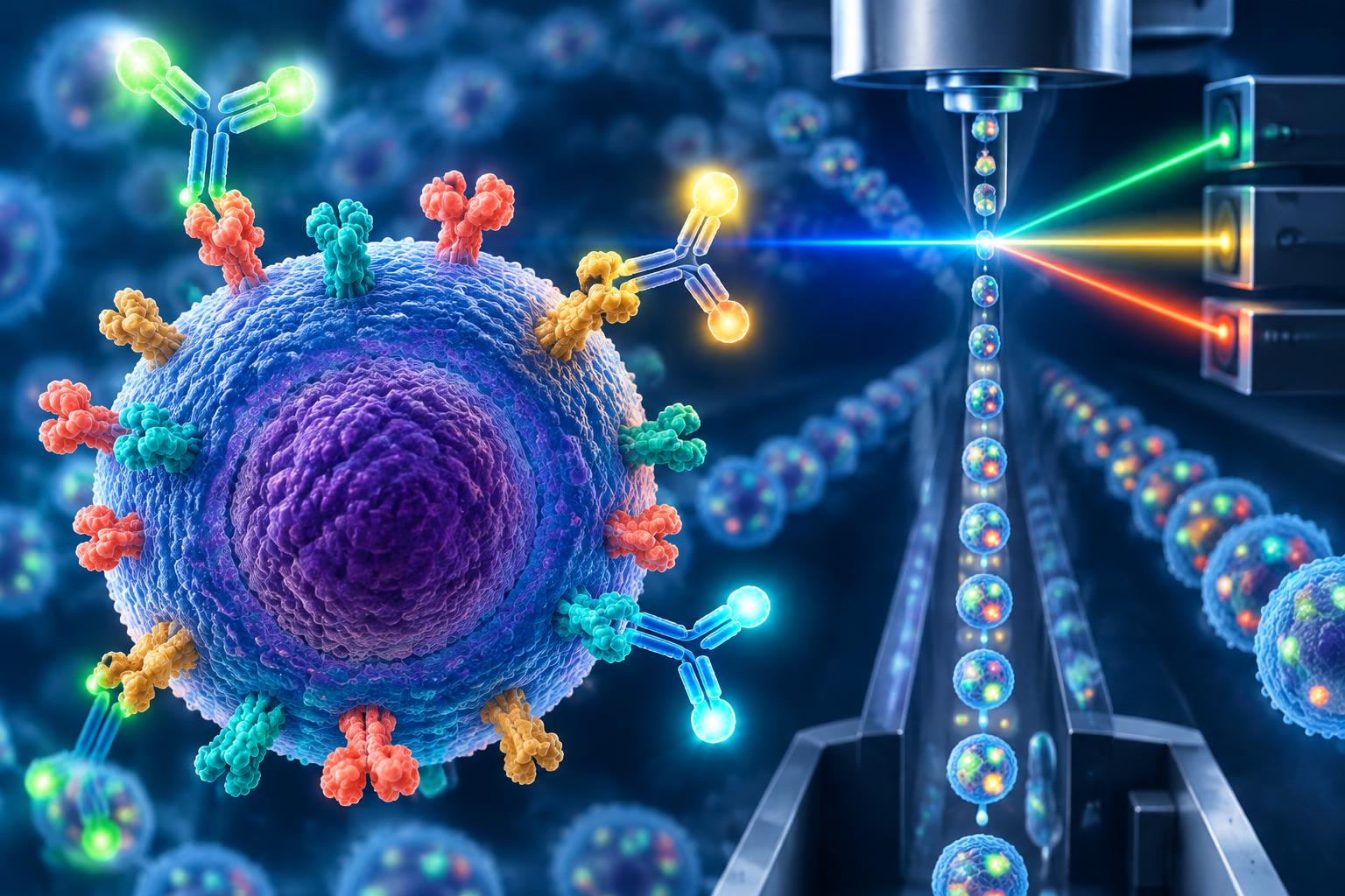
- The workhorse technology for reading a cell's marker signature is flow cytometry. Cells in a sample are tagged with antibodies engineered to bind to specific surface markers, and each antibody is linked to a different fluorescent dye.
- Why This Matters: A Real-World Example With Hematopoietic Stem Cells
- Hematopoietic stem cells (HSCs) — the blood-forming stem cells found in bone marrow, peripheral blood, and umbilical cord blood — are identified in large part by the presence of the CD34 marker on their surface. CD34 positivity has long been used as a practical, measurable proxy for isolating and counting the stem and progenitor cells responsible for reconstituting a patient's blood and immune system after a bone marrow or stem cell transplant.
- The Minimal Criteria for a Mesenchymal Stem Cell
- For mesenchymal stem cells (MSCs) — the adult stem cells most often used in orthopedic and other investigational cell-therapy research — the field's reference standard comes from a 2006 position statement by the International Society for Cellular Therapy (ISCT), commonly cited as the Dominici et al. criteria.
- Why Marker Testing Matters Beyond the Lab Bench
- Confirming a cell population's marker signature isn't just an academic exercise. It's part of how legitimate cell-therapy manufacturing verifies product identity and consistency — a necessary, though not sufficient, step toward ensuring that what's labeled as a stem cell product actually contains the cells it claims to, batch after batch.
A stem cell doesn't come with a label. Under a microscope, a mesenchymal stem cell can look a lot like an ordinary skin fibroblast, and a blood-forming stem cell looks like countless other cells circulating in bone marrow. So how do scientists actually confirm that a batch of cells is what it's claimed to be, before it's used in research or in a therapy? The answer is surface markers — proteins that sit on a cell's outer membrane and act as a kind of molecular fingerprint. This article explains what they are, how they're detected, and why a specific, internationally agreed-upon checklist of markers has become the real-world standard for defining a stem cell.
What a Surface Marker Actually Is
Every cell is studded with proteins embedded in its outer membrane, many of which project outward where they can be detected from outside the cell. Which of these proteins a cell displays, and in what combination, depends heavily on what kind of cell it is and what state it's in. Scientists have cataloged thousands of these surface proteins, many of them assigned standardized names under the "CD" system (short for "cluster of differentiation") — CD34, CD45, CD90, and so on. No single marker is unique to stem cells in general; what matters is the specific pattern, or signature, of several markers present and absent together.
How Scientists Actually Detect Them: Flow Cytometry
The workhorse technology for reading a cell's marker signature is flow cytometry. Cells in a sample are tagged with antibodies engineered to bind to specific surface markers, and each antibody is linked to a different fluorescent dye. The tagged cells are then passed single-file through a laser beam, and a detector measures which fluorescent tags light up on each individual cell. Because the test runs on thousands or millions of cells rapidly, it lets scientists determine, with real precision, what percentage of a cell population expresses a given marker — not just a yes-or-no answer, but a quantitative readout.
Why This Matters: A Real-World Example With Hematopoietic Stem Cells
Hematopoietic stem cells (HSCs) — the blood-forming stem cells found in bone marrow, peripheral blood, and umbilical cord blood — are identified in large part by the presence of the CD34 marker on their surface. CD34 positivity has long been used as a practical, measurable proxy for isolating and counting the stem and progenitor cells responsible for reconstituting a patient's blood and immune system after a bone marrow or stem cell transplant. It isn't a perfect one-to-one marker for "true" stem cells (some genuine long-term repopulating cells can be CD34-negative, and the field continues to refine purification methods), but it remains the standard, clinically actionable benchmark used to measure and release cell products for transplant.
The Minimal Criteria for a Mesenchymal Stem Cell
For mesenchymal stem cells (MSCs) — the adult stem cells most often used in orthopedic and other investigational cell-therapy research — the field's reference standard comes from a 2006 position statement by the International Society for Cellular Therapy (ISCT), commonly cited as the Dominici et al. criteria. It lays out three requirements a cell population must meet to be called an MSC for research purposes:
- Plastic adherence: the cells must stick to the surface of a standard tissue-culture flask under normal culture conditions.
- A specific surface-marker signature: at least 95% of the cell population must test positive for three markers — CD105, CD73, and CD90 — while no more than 2% may test positive for a defined negative panel: CD45, CD34, CD14 or CD11b, CD79a or CD19, and HLA-DR.
- Tri-lineage differentiation potential: under the right laboratory conditions, the cells must be able to differentiate into at least three distinct tissue types — bone-forming osteoblasts, fat-storing adipocytes, and cartilage-forming chondroblasts.
Notably, the ISCT authors themselves were explicit that these are minimal identity criteria intended for research standardization, not a clinical release specification — a distinction that matters, because a cell product being tested or sold commercially isn't automatically held to this same bar unless a specific regulatory or manufacturing standard requires it.
Why Marker Testing Matters Beyond the Lab Bench
Confirming a cell population's marker signature isn't just an academic exercise. It's part of how legitimate cell-therapy manufacturing verifies product identity and consistency — a necessary, though not sufficient, step toward ensuring that what's labeled as a stem cell product actually contains the cells it claims to, batch after batch. Without this kind of quality control, there's no reliable way to confirm that an injected product is composed of the cells its marketing describes, as opposed to a mixed population of other cell types.
Bottom Line
Surface markers give scientists an objective, measurable way to answer a deceptively hard question: what actually makes a cell a stem cell? The ISCT's minimal criteria for mesenchymal stem cells and the long-standing use of CD34 for hematopoietic stem cells are two of the field's most widely used reference standards — not because they're perfect, but because they turn an otherwise subjective judgment call into something that can be measured, repeated, and compared across different labs and manufacturers.
Key Questions Answered
- How are surface markers measured?
- Flow cytometry uses fluorescent antibody tags and lasers to quantify which markers individual cells express.
- What surface-marker pattern is required by the minimal MSC criteria?
- At least 95% must express CD105, CD73 and CD90, while no more than 2% may express the defined negative panel. Plastic adherence and tri-lineage differentiation are also required.
- Does meeting the minimal MSC criteria prove a product is clinically effective?
- No. These are research identity criteria, not a clinical release specification or proof of safety or treatment benefit.
Sources
- Minimal Criteria for Defining Multipotent Mesenchymal Stromal Cells. The International Society for Cellular Therapy Position Statement — Dominici M, et al., Cytotherapy, 2006 — https://www2.dti.ufv.br/noticia/files/anexos/phpCDlYcY_5654.pdf
- International Society for Cellular Therapy (ISCT) — Official Society Site — https://www.isctglobal.org/
- CD34 — Pathology Outlines, reference overview of CD34 as a marker of hematopoietic stem and progenitor cells — https://pathologyoutlines.com/topic/cdmarkersCD34.html
- Flow Cytometry — National Cancer Institute Dictionary of Cancer Terms — https://www.cancer.gov/publications/dictionaries/cancer-terms/def/flow-cytometry
Related Articles
- Fundamentals
Myth: No FDA Warning Letter Means a Stem Cell Clinic Is Safe and Legitimate
Why regulatory silence is not approval: warning-letter limits, the scale of unapproved stem cell marketing, a documented harm case and more meaningful checks.
- Fundamentals
Types of Chronic Pain Conditions: Which Are Being Studied With Stem Cell Therapy?
A research-based breakdown of which chronic pain conditions — from knee osteoarthritis to fibromyalgia — have real clinical trial evidence behind stem cell therapy, and which remain preclinical.
- Fundamentals
Protecting Liver Health As You Age: What the Evidence Actually Shows
How the liver ages, which habits genuinely protect it, and when fibrosis or hepatitis screening is actually recommended — evidence-based prevention, not lifestyle hype.